In general, Glaucoma occurs as a result of increased intraocular pressure (IOP) caused by a malformation or malfunction of the eyes drainage system.

Normal IOP is 19 – 21 inches of mercury.

The increased pressure causes compression of the retina and the optic nerve, and causes progressive, PERMANENT loss of eyesight if left untreated.

  • Types of Glaucoma

Glaucoma can be classified into several different and unique types:

  1. Primary Open Angle Glaucoma (POAG)
  2. Angle – closure Glaucoma (Acute Glaucoma)
  3. Congenital Glaucoma
  4. Secondary Glaucoma
  5. Pigmentary Glaucoma
  6. Normal – tension Glaucoma (NTG)

PRIMARY OPEN ANGLE GLAUCOMA

Primary Open Angle Glaucoma  is caused when the normal drainage system of the eye becomes partially blocked, causing pressure to build within the eye.  Glaucoma usually affects the perimeter vision first, with sight gradually being lost towards the center of the

Pressure builds when the drainage system is blocked.  This increasing pressure presses against the Optic Nerve and causes a gradual loss of sight.

Primary Open Angle Glaucoma

Symptoms

  • In the very early stages there are NO symptoms.
  • Mild pain in the eye, increasing gradually over time.
  • “Halos” appearing around lights.
  • Gradual loss of Peripheral vision.
  • Loss of night vision

Who is at risk?

  • Patients with a family history of Glaucoma
  • African Americans
  • Patients suffering from Myopia
  • Patients suffering from Diabetes
  • People over 35 years of age


ANGLE – CLOSURE GLAUCOMA (ACUTE GLAUCOMA)

Angle Closure Glaucoma (Sometimes referred to as Narrow Angle Glaucoma) is caused when the normal drainage system of the eye becomes suddenly blocked, causing pressure to build within the eye at a very rapid rate. Complete blindness can occur in as little as 3 to 5 days!

Pupil become more wider and blocks the passage between posterior chamber and anterior chamber.

As a result the pressure builds in posterior chamber which closes the angle in anterior chamber.

Ultimately IOP increases.                

Symptoms

  • Severe Pain
  • Pressure over the eye
  • Cloudiness to the Cornea
  • Eye extremely sensitive to light
  • Halos seen around lights.
  • Nausea and/or vomiting

Who is at risk?

  • Patients with a family history of Glaucoma
  • African Americans
  • Patients suffering from Myopia
  • Patients suffering from Diabetes
  • People over 35 years of age

CONGENITAL GLAUCOMA

Congenital Glaucoma results as a condition from birth. 

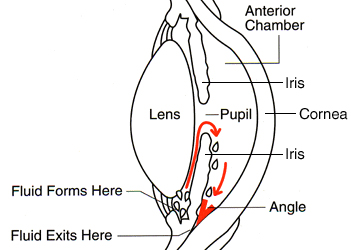
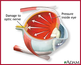
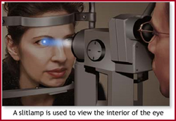
Children are born with conditions such as an abnormal development of the Anterior Chamber angles which prohibit the normal drainage of fluid from the eyes, which then causes an increase in the pressure within the eye, and subsequent Retinal and Optic Disc damage.

Parents normally are the first to recognize the symptoms of Congenital Glaucoma:

Cloudiness of the cornea due to Edema

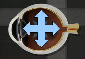
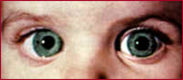
Distension (enlargement/ballooning) of the eye

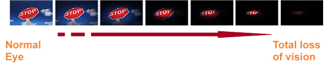
Photophobia (sensitive to light)

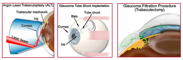
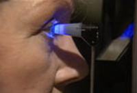
In most cases, numerous surgeries are required to correct Congenital Glaucoma. 

Lasers are sometimes used, as well as Filtration Surgery and insertion of Tube shunts:

SECONDARY GLAUCOMA

Secondary Glaucoma is usually the result of a trauma to the eye, although it can develop due to several causes:

Abnormal deposits in the eye fluid

Uveitis

Lens Changes

Drugs

Hemorrhage (escape of blood from ruptured blood vessels.

PIGMENTARY GLAUCOMA

Pigmentary Glaucoma can develop as a result of small pieces of the Iris breaking off. 

These small particles can lodge themselves in the normal drainage canals and subsequently interfere with the normal drainage of fluids from the eye.

NORMAL TENSION GLAUCOMA

Normal Tension Glaucoma occurs when there is damage to the Optic nerve detected in patients who have completely normal Inter – Ocular pressure. It has the same characteristics as Primary Open – Angle Glaucoma

Diagnosis and Treatment

Tonometry is often used as a diagnostic tool.  The Tonometer is gently pressed against the eyeball, and the resistance (internal pressure) is measured.

Gonioscopy can be used to determine if the angle where the iris meets the cornea is open or closed.

Perimetry is an essential method used to determine if there is any loss of the visual field.

Slit Lamp Examination is another method of diagnosis of patients with suspected Glaucoma.

Stereo disc photography of the optic disc is performed to determine if there is abnormal cupping in the optic nerve head. Subsequent exams or photos are compared over time.

Treatment

  • Medication
  • Laser Surgery
  • Filtration Surgery